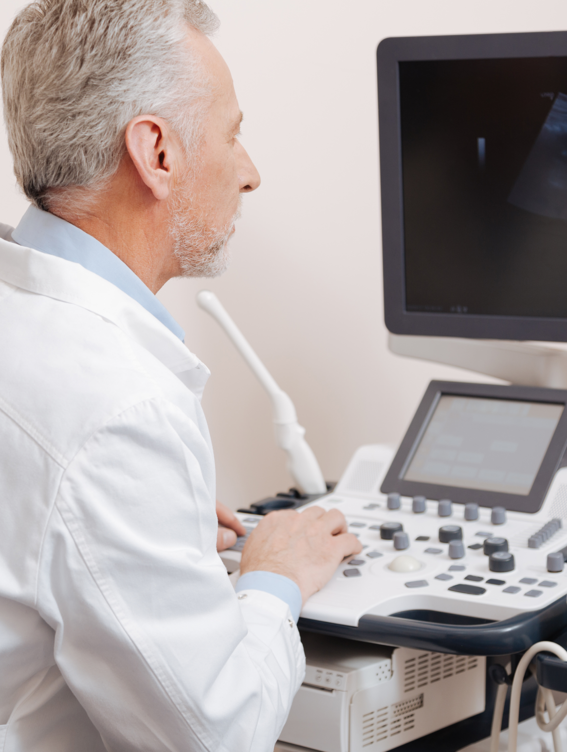
Ultrasound Imaging
It is a uses ultrasound waves, and it is commonly used to monitor fetal health during pregnancy, but it can also be used to diagnose other internal organs, such as the glands, kidneys, liver, intestines, and other vital organs, as well as blood vessels and the heart.
Our clinic provides accurate ultrasound imaging due to the use of state-of-the-art diagnostic devices such as Samsung HS30, which comes with improved tools to help you provide better services for your patients through advanced imaging techniques such as:
1- ClearVision
The noise reduction filter improves edge enhancement and creates sharp 2D images. ClearVision provides application-specific optimization and temporal resolution in live scan mode.
2- Knee with ClearVision
S-Harmonic™ using pulse inversion technology improves image clarity, near to far. Reducing signal noise, S-Harmonic™ provides more uniform ultrasound images.
3- S-Flow™
S-Flow™, a directional Power Doppler imaging technology, can help to detect even the peripheral blood vessels.
4- ElastoScan™
A diagnostic ultrasound technique for imaging elasticity, ElastoScan™ detects the presence of solid masses in tissues and converts any stiffness into color images.
5- AutoIMT+
AutoIMT+ is a screening tool to analyze a patient’s potential risk of cardiovascular disease. It allows easy intima-media thickness measurement of both the anterior and posterior wall of the common carotid by the click of a button. This simple procedure enhances exam productivity and adds diagnostic value.
6- Strain+
Strain+ is a quantitative tool for global and segmental wall motion of the left ventricle (LV). In Strain+, three standard LV views and a Bull’s Eye are displayed in a quad screen for an assessment of the LV-function.
7- Panoramic
Panoramic imaging displays an extended field-of-view allowing users to examine a wider area. Panoramic imaging also supports angular scanning with data acquired from the linear and convex transducer.
8- NeedleMate+™
NeedleMate+™ helps needle targeting when performing commonly used intervention procedures.
Book an appointment and find out more about our clinic’s HS30 device and how it can make a difference to your diagnosis!!

Ultrasound Imaging Procedure:
The patient is placed in a comfortable position for the imaging. A special device is used to emit ultrasound waves towards the area of the body to be imaged, and the returning waves are analyzed immediately. The radiologist sees the image and diagnoses it. The radiologist takes images to illustrate the necessary scenes for the treating physician, which are attached to the final report.

