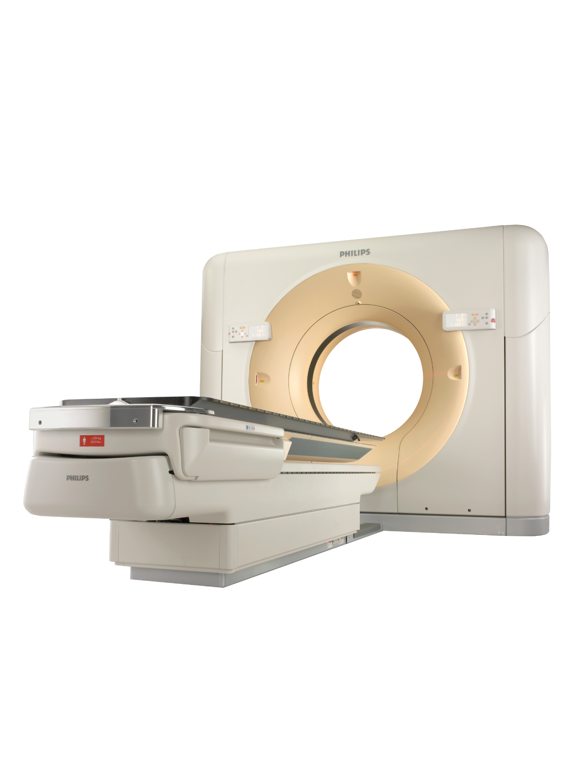
Multi-slice CT imaging technology
This technology uses X-rays to produce three-dimensional internal images of different parts and slices of the body. It is used in the diagnosis of malignant and benign tumors in multiple areas of the body, brain imaging, internal bleeding, calcifications, and abdominal and pelvic imaging for various diagnoses.

CT imaging procedure:
The imaging process may take about 10 to 20 minutes.
- The imaging does not affect dental fillings or props.
- The patient takes off the jewelry he wears before the imaging because they will affect the results of the imaging.
- The patient is prepared for the imaging and may need to undergo certain laboratory tests before the opaque material is injected.
- The patient is placed in the machine and a thin beam of X-rays is rotated around the patient's body.
- The X-rays are recorded and converted into three-dimensional images by a computer.
- Pregnant women are not allowed in the imaging room to protect the fetus from exposure to X-rays.

