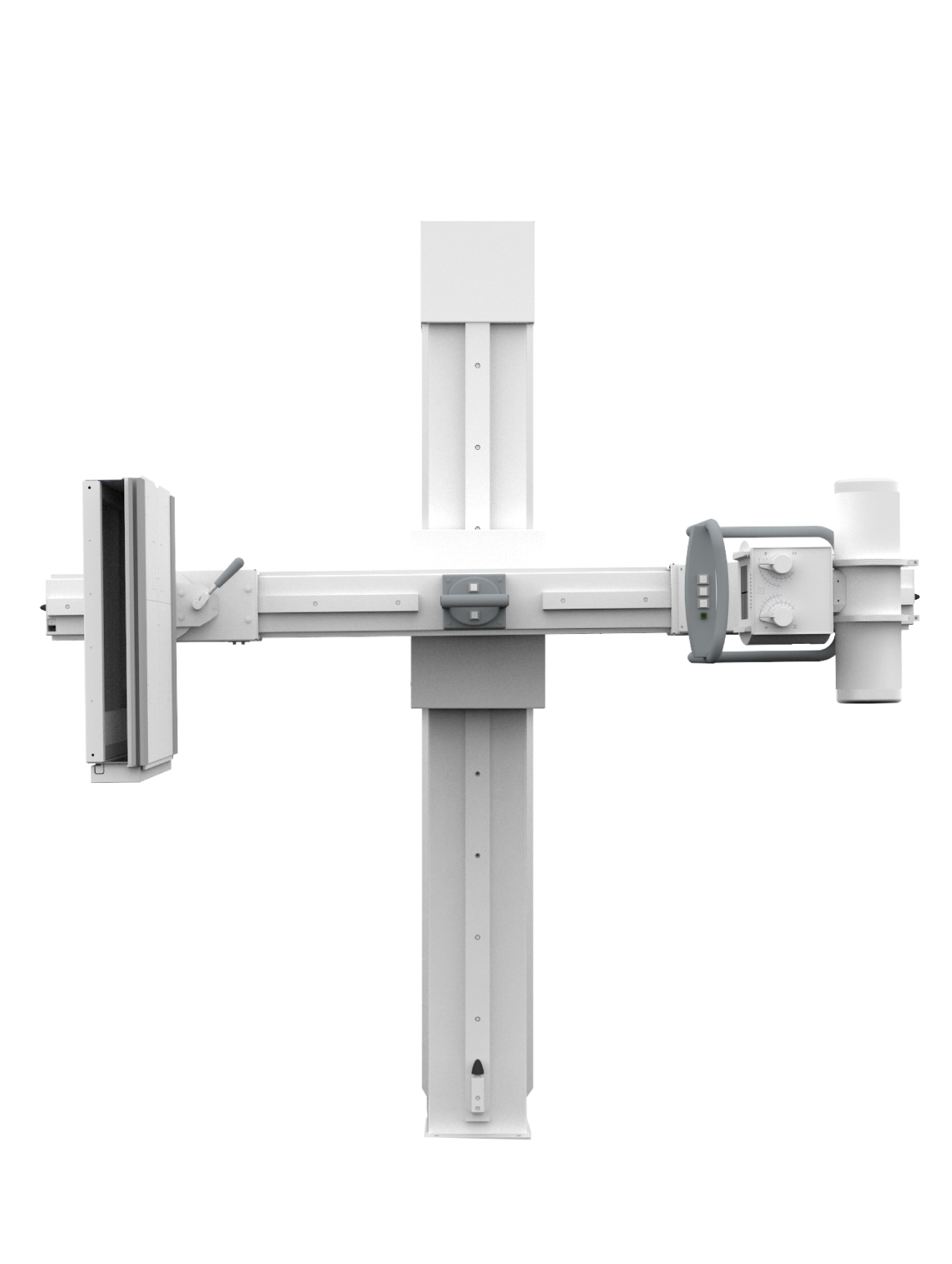
Contrast Radiography
Contrast radiography is a medical technique that involves the use of X-rays and a special dye called a contrast medium to examine the organs. This method enables the radiologist to study structures that are not visible in normal X-ray exams.
X-rays work by passing through the body. Bones are easily visible on X-rays because they block the rays. But organs, blood vessels, the stomach, and the colon are not as dense as bones, and therefore, they don’t block the X-rays as effectively. The contrast medium is used to highlight these specific areas in the body, making them more visible and detailed in the X-ray image.

There are several types of contrast imaging, including the following:
- Ascending Descending Urography.
- Urethrography.
- Gastrophotography.
- Gastrointestinal Contrast Imaging.
- Colonography.
- T-tube cholangiography.
- Hysterosalpingography to study female infertility by professional female doctors.
- External Tube Imaging.
- Distal Colostography.
- Enma Contrast Imaging.

